Wednesday, September 30, 2009

Stomach Cancer

Stomach cancer is one of the common cancers. It is more prevalent in Japan & South America. The incidence increases with age and there is a slight male preponderence.


Stomach Cancer as seen thru an OGDS

The risk factors for stomach cancer include:
-H.Pylori infection
-smoked food
-preservatives/high salt diet
-smoking/alcohol consumption
-pernicious anaemia
-previous gastrectomy (stomach surgery)
-stomach polyps
-genetics: blood group A, hypogammaglobulinaemia

Patients who have stomach cancer may present with:
-upper abdominal pain
-loss of weight
-loss of appetite
-early satiety
-persistent vomiting
-vomiting blood (haematemesis)
-passing blackish stools (malaena)
-symptoms of gastritis such as dyspepsia, belching

Or they may be asymptomatic.

To confirm the diagnosis, an Oesophago-Gastro-Duodeno-Scopy (OGDS/EGDS) should be done. The OGDS will be able to visualise the tumour and assess the size and also stop any bleeding. A biopsy from the tumour can be obtained with the OGDS to confirm the diagnosis.

To properly assess the extent of spread of the cancer, the patient should undergo a CT-scan or MRI or PET scan. Endoscopic ultrasound is able to assess the depth of the tumour. And staging Laparoscopy will be able to properly stage the cancer and assess for peritoneal deposits.


Fungating Stomach cancer noted in a resected specimen

The cure for stomach cancer is surgery. A subtotal or total gastrectomy (surgical excision of the stomach) can be done. The type of surgery will depend on the stage of the cancer and also the patient's fitness for surgery. Curative surgery (D2 classification) includes surgical excision of the stomach with a clear micro & macroscopic margin, removal of lymph nodes one level beyond those involved &  removal of the omentum, spleen and distal pancreas.

Currently, based on the MAGIC Trial, a 3 cycle neoadjuvant (pre-operative) chemotherapy is recommended followed by surgery and another 3 cycles post-surgery. This has been shown to downstage the cancer, improve resectability and improve overall survival for the patient.

Tuesday, September 22, 2009

Peptic Ulcer Disease (Stomach Ulcers)

Peptic ulcer disease is common. Patients usually complain of upper abdominal pain, dyspepsia, water-brash, chest pain, difficulty breathing, belching and feeling of bloatedness. Peptic ulcer disease comprise of gastritis and stomach or duodenal ulcers.


The main causes are:
-ingestion of NSAIDs such as aspirin, voltaren, ponstel/ponstan (mefenemic acid)
-H. Pylori infection
-stress
-stomach cancer or lymphoma
-hiatus hernia
-hyperparathyroidism
-Zollinger-Ellison syndrome (a tumour of the pancreas which produces the hormone gastrin which increases stomach acid secretion)

The development of peptic ulcers is due to the imbalance between protective factors and aggressive factors. Protective factors include mucin, bicarbonate and prostaglandins which are naturally secreted by the cells from the stomach. Aggressive factors include H. pylori, NSAIDs and excessive acid secretion from the stomach.


Stomach ulcer

Helicobacter pylori is a bacteria which causes inflammation of the stomach mucosa. Certain strains are more virulent such as the CaG and VacA which promotes the release of pro-inflammatory substances in the stomach. H. pylori infection is also a risk factor for stomach cancer. It is classified as a carcinogen.

NSAIDs cause peptic ulcers by reducing the secretion of protective prostaglandins from the stomach. It is a known side effect of the drug due to inhibition of cyclo-oxygenase (COX). The newer selective COX-2 inhibitor NSAIDs (eg celecoxib, etoricoxib) causes less gastric complications.

Patients who are <55 years old and with symptoms of peptic ulcer disease can undergo non-invasive investigations such as:
- Urea breath test: 95% sensitive in detecting H. pylori infection
- H. pylori serology blood test: >80% sensitive in detecting the infection
- Stool H. pylori antigen test: >90% sensitive in detecting the infection

Patients who are >55 years old or have "Alarm Symptoms" should undergo an urgent OesophagoGastroDuodenoScopy (OGDS/EGDS).
Alarm Symptoms include:
- weight loss
- difficulty swallowing
- vomiting blood or passing blackish stools
- anaemia
- a palpable mass in the upper abdomen
- persistent vomiting


OGDS/EGDS: the fibre-optic scope

OGDS/EGDS is a simple day-care procedure where a small fibre-optic scope is passed thru the patients' mouth into the stomach to visualise the oesophagus, stomach and duodenum. It can be done with or without sedation. The advantages of OGDS is that it is able to diagnose the disease and also localise it. Pictures can be taken of the ulcer areas to compare after treatment. Biopsies can be taken for H.pylori hustology and also for suspicious stomach tumours.


View from OGDS of a normal stomach at the pylorus

The treatment for mild gastritis includes:
-dietary advice to avoid sour & spicy foods
-reduce alcohol and stop smoking
-antacids
-H2-blockers such as ranitidine tablets

Patients with H. pylori infection should be treated with a combination of antinbiotics and proton-pump inhibitors (PPI). The first line therapy include:
-Clarithromycin + Amoxycillin + PPI (pentoprazole/esomeprazole) for 2 weeks

Patients who are allergic to penicillin should be treated with:
-Clarithromycin + Metronidazole + PPI

Patients who do not respond to first line therapy should be treated with:
-PPI + Bismuth + Tetracycline + Metronidazole

Post treatment, the patients should get a repeat Urea Breath Test to confirm that the infection has resolved. And these patients should be continued on PPIs for another 1 - 2 months.  

Complications of peptic ulcers include:
-Perforated gastric ulcer
-Bleeding from the ulcers
-Erosion and penetration into other organs
-Gastric outlet obstruction


Friday, September 18, 2009

Acute Pancreatitis

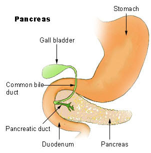
Pancreatitis is the inflammation of the pancreas. The pancreas is an organ anatomically located just underneath our stomach which helps to digest food and also secretes hormones. Among the important hormones secreted are insulin and glucagon which helps to regulate our blood sugar.


Pancreatitis can be divided into acute or chronic pancreatitis depending on the duration and onset of the inflammation.

Patients with Acute Pancreatitis usually present with severe upper abdominal pain which radiates to the back, nausea & vomiting with a history of gallstones or alcohol bingeing. The causes of acute pancreatitis include:
- gallstones
- alcohol ingestion
- trauma (blunt or penetrating) to the abdomen
- post abdominal surgery or ERCP (endoscopic retrograde cholangiopancreaticogram)
- pancreatic tumours or structural abnormalities
- medications such as corticosteroids, diuretics, ACE inhibitors
- autoimmune pancreatitis
- viral infection such as mumps
- high calcium levels
- high fat (triglyceride) levels

Investigations for patients with Acute Pancreatitis include:
- raised serum amylase (usually taken as >4 times normal lab value)
- raised serum lipase
- raised urinary amylase
- raised C-reactive protein
- radiological investigations such as ultrasound or CT scan to look for gallstones or other abnormalities

Patients who are diagnosed with Acute Pancreatitis will need to be admitted into the hospital for close observation. This is because the disease might worsen and lead to severe sepsis and multi-organ failure. The severity of the disease is classified according to the Imrie-Glasgow, Ranson or APACHE II criterias.
Patients are usually kept fasted to reduce the pancreatic function and started on intravenous fluids and also analgesics. They are also given supplemental oxygen.
Patients who have Acute Pancreatitis due to gallstones will need a cholecystectomy (surgical removal of the gallbladder) to avoid further recurrence of the disease.
Patients who are chronic alcoholics should be referred for counselling and also taught coping strategies and encouraged to join Alcoholic Anonymous and other support networks.

The sequelae of Acute Pancreatits include:
- complete recovery (majority of patients)
- destruction and infection of part of the pancreas (necrosis)
- acute fluid collection around the pancreas
- pancreatic pseudocyst (collection of fluid around the pancreas seen >4 weeks after the initial diagnosis)
- pancreatic abscess

Saturday, September 12, 2009

Gallstones

Gallstone disease is common; affecting around 80% of the population. However, of all the patients who have gallstones, only 20% are symptomatic.

There are 3 main types of gallstones:

- Cholesterol stones (80%): commonly occurs in patients who are obese or have high cholesterol and lipids.

- Pigmented stones: Black stones – associated with blood disorders (haemolysis), Brown stones – due to infection of the biliary tract.

- Mixed stones: cholesterol & pigmented types.


Risk factors for gallstones include:

- hereditary

- female sex

- obesity

- high cholesterol levels

- diabetes

- increasing age

- oral contraceptive pills

- post bariartric (obesity) surgery

- inflammatory bowel disease

- haemolytic anaemia


Patients with gallstones can present with:

- abdominal pain usually at the right upper quadrant (biliary colic)

- inflammation of the gallbladder (acute cholecystitis)

- swelling of the gallbladder due to collection of mucous (mucocoele)

- infection of the gallbladder (empyema)

- perforation of the gallbladder (gangrene & perforation)

- jaundice due to obstruction of the bile duct (Mirizzi’s Syndrome)

The gallstones can also migrate into the bile duct & cause obstruction, jaundice & infection.

Common investigations to diagnose gallstones include:

- ultrasound of the abdomen

- CT/MRI scan of the abdomen

- Endoscopic Retrograde Cholangio Pancreaticogram (ERCP): to assess the biliary tract and to remove any stones in the biliary tract.

Treatment:
- Surgery to remove the gallbladder (Cholecystectomy): usually done laparoscopically but can also be done as an open surgery in complicated cases.
- Patients with Acute Cholecystitis will have to be admitted to the hospital for intravenous antibiotics.

- Patients with empyema, gangrenous or perforated gallbladder will need emergency surgery to remove the gallbladder.

Wednesday, September 2, 2009

Thyroglossal Cysts


A Thyroglossal Cyst is a remnant of the thyroglossal tract which connects our tongue to the thyroid gland. During its formation, the thyroid gland bud migrates from the base of our tongue (foramen caecum) to our lower neck. It travels along the thyroglossal tract. The Thyroglossal cyst forms due to the persistent remnant of the thyroglossal tract. Usually the tract disappears completely after the thyroid gland is formed. The tract is closely related to the hyoid bone (a semi-circular bone located just above the thyroid cartilage).
Thyroglossal cysts usually present as a swelling at the middle part of the neck above the thyroid cartilage (adam's apple). However, it can also be located anywhere from the base of the tongue up to just above the sternum (chest bone). It is usually painless but sometimes can get infected. When it gets infected, it might be inflammed & painful. Occasionally the infected thyroglossal cyst might rupture and form a thyroglossal sinus.

It is important to confirm that the swelling is not a lingual thyroid gland prior to surgery. This can be done with an ultrasound scan or a thyroid radioisotope scan.
The treatment for thyroglossal cyst is surgery. This surgery is called Sistrunk's surgery. It involves making a small incision on the cyst and removing it completely together with the remnant tract and also part of the hyoid bone. It is not a major surgery but as always all surgery will carry some risks.

Infected thyroglossal cysts should not be operated on. It should be treated with antibiotics first and planned for a surgery later when the infection subsides.

There is a small risk of malignancy (papillary carcinoma) with the thyroglossal cyst although it is very rare.

Differential diagnosis for a thyroglossal cyst include dermoid cyst, sebaceous cyst, lipoma, lingual thyroid gland and lymph nodes.