Tuesday, December 15, 2009

Colon Cancer

Colon cancer is a malignant tumour involving the large intestines. It is one of the more common cancers in our society. The commonest type of colon cancer are adenocarcinomas (95%) with the remaining 5% comprising of mesenchymal tumours (GIST), neuroendocrine tumours, lymphomas and sarcomas.


Surgical specimen of colon cancer with polyps

The risk factors include:
- older age
- diet high in fat and low in fibre
- hereditary (20% of cases including Familial Adenomatous Polyposis & Hereditary Non-Polyposis Colon Cancer)
- male sex
- patients with inflammatory bowel disease (especially ulcerative colitis)
- colonic polyps
- obesity
- cigarette smoking
- acromegaly
- patients with an uretero-sigmoidostomy

Patients usually present with:
- altered bowel habits: constipation alternating with bouts of foul smelling watery stools
- bloody stools
- mucous in the stools
- loss of weight and loss of appetite
- incidental finding on screening colonoscopy
- symptoms of metastasis (malignant spread) eg difficulty breathing, liver swelling, abdominal distension, bone pain, confusion, weakness

Patients can also present as an emergency case with:
- intestinal obstruction with or without abdominal distension
- perforation of the large intestines
- symptoms of anaemia due to profuse bleeding from the tumour

Investigations include:
- colonoscopy which can visualise and biopsy the tumour
- CT-scan to look for spread (metastasis) of the tumour to the lungs, liver or lymph nodes
- serum CEA levels (tumour marker for colon cancer): raised CEA reinforces the diagnosis and can be used for surveillance to detect recurrence
- genetic testing for hereditary colon cancers eg APC, hMLH1, hMSH2, hPMS1 genes


Colonoscopy image of colon cancer

Management of patients with colon cancer will depend on the stage and extent of the disease. The mainstay of treatment for colon cancer is surgery. The type of surgery will depend on the site of the cancer. A right hemicolectomy is done for cancers at the right side of the colon. Left hemicolectomy for cancers of the left colon, anterior resection for cancers of the sigmoid and rectum.


Patients with advanced stage 2 and stage 3 colon cancers will require chemotherapy after surgery. Common chemotherapy regimes used include 5-FluoroUracil, Oxaliplatin and Irinotecan. Radiotherapy is indicated when surgical margins are involved. Newer treatment options include the use of molecular targeted therapy such as bevacizumab and cetuximab.   
Patients who present with metastatic disease (stage 4) will usually be treated with palliative and supportive care. However, recent advances have shown that resection of metastatic liver and lung lesions improves patient survival.

Tuesday, November 17, 2009

Acute Appendicitis

The appendix is a tubular, blind-ended, vestigial structure which is attached to the caecum. Anatomically the location of the appendix corresponds to the right iliac fossa which is at the lower right quadrant of our abdomen. The human appendix is hypothesised to have lost its original function through evolution.


 Some of the functions of the appendix are:
-immunological functions - it contains lymphoid follicles which secrete immunoglobulins
-it contains serotonins which are neuroendocrine hormones
-it has a bacteriological function where it helps to control the normal gut bacterias
-Surgical functions - where the appendix is used as an 'urinary conduit' in bladder reconstruction in bladder surgery & it can also be used as an 'appendicostomy' which can be used for antegrade colonic enema (wash-out of faeces) in patients with constipation.

The appendix lies in close proximity to the caecum (the beginning of our large intestines). Approximately 75% of the appendix lies behind the caecum (retrocaecal), 20% in the pelvis and the remaining 5% either pre- or post-ileal (infront or behind the small bowel).


Appendix attached to the caecum

Acute appendicitis is the inflammation of the appendix. It is the commonest acute surgical condition in the abdomen. It typically presents with an initial pain around the umbilical region which later migrates and localises in the right iliac fossa. Usually the patient will have a low grade fever, loss of appetite and nausea. There will be tenderness of the abdomen localised at the right iliac fossa which is typically described as at McBurney's point (2/3 along the line drawn between the umbilicus and the anterior superior iliac spine).
Severe cases of appenditis can present with severe abdominal pain, high spiking temperature & dehydration. This usually happens when there is a delay in diagnosis and the appendix perforates leading to purulent or faeculent peritonitis (contamination of the abdominal cavity with pus or faeces).

Other causes of a right iliac fossa pain are:
-urinary tract infection
-kidney stones (renal colic)
-Meckel's diverticulitis
-mesenteric adenitis (usually associated with flu)
-colonis diverticulitis (usually in elderly people)
-torsion of ovary
-ectopic pregnancy
-pelvic inflammatory disease
-intestinal obstruction

The treatment for acute appendicitis is appendicectomy (surgical excision of the appendix). This procedure was traditionally done as an open surgery but most surgeons are doing laparoscopic (minimally invasive) appendicectomies now. The complications of appendicetomy includes:
-wound infection (<5% risk)
-intra-abdominal abscess
-faecal fistula (leakage of faeces from the resected appendix stump)

The sequelae of acute appendicitis includes:
- Perforated appendix: - as a result of inflammation and ischaemia (reduced blood supply) to the appendix, the wall weakens and perforates. This will lead to localised or generalised, purulent or feaculent peritonitis. The patient will be more ill.
- Appendicular mass: - a swelling noted at the right iliac fossa which is a mass of omentum covering the inflammed appendix. This typically happens a few days after the initial episode of right iliac fossa pain when our body tries to wall of the inflammed appendix.
- Chronic appendicitis: - this usually occurs in patients who were treated with antibiotics or anti-inflammatories for a bout of lower abdominal pain. They develop a chronic grumbling right iliac fossa pain which is very vague. It is usully a diagnosis of exclusion.

Tuesday, November 3, 2009

Abdominal Hernias

Hernias are abnormal protrusion of organs from the wall which contains it. Abdominal hernias are usually inguinal hernias, femoral hernias, paraumbilical hernias or incisional hernias.
Inguinal & femoral hernias occur in the inguinal (groin) region and may occur on one side (unilateral) or both sides (bilateral).


A huge Right sided indirect inguinal hernia which has entered the scrotum

Inguinal hernias are the commonest type of abdominal hernias. The main causes are: chronic cough, straining due to constipation or urine obstruction, weakness of the abdominal muscles or congenital defects (patent processus vaginalis). Inguinal hernias can be further divided into direct or indirect inguinal hernias but the management for both is the same. Direct inguinal hernia occurs thru the weakness in the transversalis muscle of the abdominal wall while indirect inguinal hernia occurs thru a defect in the deep inguinal ring. 
Both present with a swelling in the groin which expands on standing or upon straining. Most of the time the swelling is reducible. Sometimes the hernia may be stuck (incarcerated) or obstructed or strangulated (blood supply compromised). The swelling in an indirect inguinal hernia may enter into the scrotum in men. 
The management for inguinal hernia include treatment of the causes of the hernia and the mainstay of treatment is surgery.
Surgery can be either open surgery or laparoscopic surgery. Open surgery is a simple procedure which can be done under local anaesthesia as a day case in uncomplicated hernias. The hernia is reduced, the hernial sac excised and a polypropylene mesh inserted above the muscles to strenghten it. This is known as the Lichtenstein repair. Complicated cases (incarcerated or obstructed or strangulated) would need general anaesthesia and hospital stay. At the moment laparoscopic is indicated for recurrent hernias or bilateral inguinal hernias. The scars are smaller, it is less painful and patients are discharged earlier. 

Femoral hernias occur thru the femoral canal in the groin. The presentation is the same as inguinal hernias with a groin swelling. However, it has a much higher risk of strangulation due to the narrow opening of the femoral canal. Femoral hernias are more common in women. Treatment is with surgical repair which can be done under local anaesthesia for uncomplicated cases. Some surgeons approximate the femoral canal with sutures and reinforce with a pectineal fascia flap while some use a polypropylene mesh plug repair. Laparoscpoic repair can also be done. 

Paraumbilical hernias occur commonly is people with ascites (excess fluid collection in the abdominal cavity), those who are obese and those who have abdominal wall weakness. The swelling is usually noted just around the umbilicus. This should not be confused with umbilical hernias which occur in babies due to congenital defects. Paraumbilical hernias are repaired surgically. The surgery entails reduction of the hernial contents, excision of the hernia sac and application of a big piece of polypropylene mesh to cover the defect. 

Incisiona hernias occur as a late complication of abdominal surgery. A history of infection of the abdominal wound, malnutrition, complicated bowel surgery and obesity are predisposing causes. The swelling occur just under the incision scar of the abdominal wound. Surgery includes revision of the previous scar, reduction of the hernial contents and excision of the hernial sac. A big polypropylene mesh is used to cover the defect. Laparoscopic repair is also feasible for both paraumbilical and incisional hernias.

The complications of hernial repair are: bruising, bleeding, infection of the wound, infection of the mesh, seroma collection and recurrence. 
Complications specific to inguinal hernia repair include: ischeamic orchitis, testicular atrophy, scrotal oedema, hydrocoele, chronic pain due to neuroma or osteitis and numbness around the groin area.      

Thursday, October 15, 2009

Breast Cancer

Breast cancer is the commonest cancer in women all over the world. The incidence is highest in women around the age of 40 - 60 years but it may also occur at any age. The lifetime risk of developing a breast cancer in a woman is 11%.

Patients with breast cancer commonly present with:
- breast lump
- retracted nipple
- lymph node swelling in the axilla (armpits)
- ulcerated swelling on the breast
- redness & inflammation of the breast
- nipple discharge which can be blood or mucous
- incidental finding on screening by mammogram

95% of breast cancer occur sporadically while only 5% are due to genetic factors. The genes which are implicated in breast cancer are BRCA-1 and BRCA-2. Patients with BRCA genes have a 50% lifetime risk of breast cancer and a 30% risk of getting a breast cancer on the opposite breast. Women with BRCA-1 also have a 30% risk of getting ovarian cancer.

Other genetic syndromes associated with breast cancer are:
- Li Fraumeni syndrome which includes brain tumours, sarcomas and leukaemias
- Cowden syndrome which includes thyroid tumours and skin lesions
- Peutz-Jaghers syndrome which includes gastro-intestinal tract polyps, pigmentation of the lips and increased risk of gastro-intestinal cancers.

To confirm the diagnosis of breast cancer, 'Triple Assesment' is recommended. This includes:
- breast examination by the doctor
- tissue biopsy with Fine Needle Aspiration Cytology (FNAC) or Core Biopsy
- radiological investigations with a Mammogram or Ultrasound or MRI


Mammogram

Patients with strong family history of breast cancer should undergo genetic testing for BRCA-1 and BRCA-2 genes.

Once the diagnosis of breast cancer is confirmed, a patient is counselled on her diagnosis and the plan of management. The management will depend on the stage of the cancer, type of cancer (DCIS vs Invasive Ductal/Lobular Cancer) and patient's Estrogen/Progestrone Receptor (ER/PR) status.

For patients with non-metastatic (have not spread beyond the breast) cancer, the initial treatment is mastectomy (breast removing surgery) with removal of lymph nodes. The indications for mastectomy are:
- Invasive breast cancer more than 3cm
- multifocal DCIS
- patients with recurrent cancer after breast conserving surgery
- young patients who have a high risk of cancer recurrence (BRCA carriers)
- old patients who do not want to undergo further surgery
- male patients with breast cancer

Patients with DCIS (ductal carcinoma in situ) or Invasive breast cancer less than 3cm can undergo Breast Conserving Surgery (BCS) with post-operative radiotherapy. However, there is a 10% chance that further surgery might be required with BCS.

Removal of lymph node is needed for control of loco-regional disease and for more accurate staging of the disease. A few techniques commonly used are lymph node dissection or sampling or Sentinel Lymph Node Biopsy.

Post mastectomy, if the patient has got lymph node involvement or the tumour is bigger than 1.5cm, it is necessary for the patient to undergo chemotherapy which will further destroy the cancer cells in the body. The common regime used is a combination of 5-FluoroUracil, Epirubicin and Cyclophosphamide.

Patients who have ER/PR positive should be started on Tamoxifen tablets (20mg daily) which will reduce the level of estrogen in the body and reduce the risk of recurrence of the cancer. Post menopausal patients can be started on aromatase inhibitors such as anastrozole or letrozole which also reduces estrogen levels.

Patients who are HER-2 (human epidermal growth factor receptor-2) positive can be started on trastuzumab which is shown to improve disease  free survival. However, this drug is contraindicated in women with heart failure.

Patients with locally advanced breast cancer (size more than 5cm or with involvement of chest wall or skin) should undergo pre-operative chemotherapy to reduce the size of the cancer. The regime is the same as the post-operative one or a taxane based (docetaxel) chemotherapy can also be used.

Post-mastectomy, patients who are keen for reconstruction of their breast should be referred to a Plastic Surgeon for reconstructive surgery. The breast can be reconstructed with the patients own muscles and skin (free or pedicled flap reconstruction) or with a prosthetic breast implant.

Monday, October 12, 2009

What is a Cancer?

A Cancer is defined as an abnormal mass of tissue, growth of which is uncoordinated and exceeds that of normal tissue which persists even after cessation of the stimuli which provoked it. The main hallmarks of cancer are that it is purposeless, it preys on its host and it is autonomous.

The biology of Cancer growth are:
- Self sustained growth signals
- Insensitivity to inhibitory growth signals: Normal cells respond tumour suppressor genes.
- Limitless growth potential: It does not stop growing unlike normal cells.
- Evasion of apoptosis (programmed cell death): Normal cells have a fixed life-span.
- Ability to metastasise and invade
- Ability to produce own blood supply (angiogenesis)

The common causes of cancer can be divided into:
- Radiation eg UV light, ionising radiation, nuclear radiation exposure
- Infection eg H.Pylori (stomach cancer), schistosomiasis (bladder cancer), Hepatitis B & C (liver cancer)
- Genetic abnormalites eg Familial Adenomatous Polyposis, Li Fraumeni Syndrome
- Chemicals eg aniline dye (bladder cancer), cigarette smoking
- Others eg diet, chronic irritation (Marjolin's ulcer)

Treatment options for Cancers include:
- Radiotherapy
- Chemotherapy
- Surgery
- Hormonal Therapy
- Molecular Targeted Therapy

Tuesday, October 6, 2009

Deep Venous Thrombosis

Deep Venous Thrombosis (DVT) is a commonly discussed medical condition. It is a condition where a blood clot forms in the deep venous system of the lower legs. DVT is the third commonest cardiovascular disease. Around 20% of DVTs occur post surgery. 25% of DVTs are associated with cancers and 50% happen due to unknown reasons.


Anatomy of Deep Venous System

The aetiology of DVT is summarised by Virchow's triad of:
-Hypercoagulability (increased tendency to form blood clots)
-Venous Flow Stasis
-Vessel wall damage


Patients who have DVT usually complain of:
-calf pain
-calf swelling
-redness around the lower leg
-unilateral pitting oedema
-low grade fever


Sometimes the lower leg can be bluish (cyanotic), severely painful and swollen in a condition described as Phlegmasia Caerulia Dolens. Phlegmasia Alba Dolens describes a pale, painful, swollen limb.


Conditions which increase the risk of DVT are:
-increasing age
-immobility
-pregnancy
-patients who are diagnosed with cancers
-patients who are on oral contraceptive pills (combined progestrone & oestrogen)
-patients who are on hormone replacement therapy
-obesity
-patients with inflammatory bowel disease
-patients who are planned for knee, pelvic & hip surgeries
-patients with heart failure


Investigations for patients with DVT include:
-Duplex ultrasound scans: - 95% sentitive & specific
-Venography: - can be both diagnostic & therapeutic
-Magnetic Resonance Venography
-Impedence Pletysmography
-Spiral CT-scan / CT Pulmonary Angiography: to rule out Pulmonary Embolism


Image at Left: Deep Vein Thrombosis
Image at Right: Embolus propagating

Treatment for DVT aims to stop further clot formation, to break down the existent clot (fibrinolysis) and to prevent pulmonary embolism. Pulmonary embolism is the propagation of a blood clot from the lower leg into the smaller veins of the lungs. This is a potentially deadly complication of DVT where 10% of patients with symptomatic pulmonary embolism die within the first hour. 


Treatment options include:
-intravenous Heparin infusion
-oral Warfarin tablets
-subcutaneous Low Molecular Weight Heparin (eg enoxaparine) injection
-percutaneous mechanical thrombectomy
-catheter directed thrombolysis
-insertion of an Inferior Vena Cava filter: for prevention of pulmonary embolism in high risk patients


The complications of DVT are:
-pulmonary embolism
-propagation of clot
-chronic venous insufficiency
-varicose veins
-venous ulcers

Sunday, October 4, 2009

Varicose Veins

Varicose veins are common among the general population. It is defined as tortuous, dilated, abnormal veins of the lower limbs. The other variants of these unsightly veins are reticular veins (superficial, prominent veins) and thread veins (fine, thin cutaneous veins).


Varicose veins are more common in women, caucasians and in those with a family history. The risk factors are:
-obesity
-pregnancy
-prolonged stationary standing
-women taking oral contraceptive pills or hormone replacement therapy

The aetiology of varicose veins can be due to:
-venous valve insufficiency which is either congenital or acquired. Congenital insufficiency occurs in patients who are born with weakness of the venous valves. Acquired insufficiency is secondary to thrombosis (blood clots) occuring around the valves.
-perforator (the connecting veins between the deep venous & superficial venous systems) insufficiency
-disturbance to venous flow such as deep venous thrombosis (DVT), pelvic vein thrombosis and vascular malformations.

Patients who have varicose veins usually present with:
-unsightly dilated veins
-itchiness around the veins
-aching sensation of the lower llimbs
-swelling of the lower limbs
-throbbing pain of the lower limbs

The complications of varicose veins are:
-bleeding (usually due to accidental trauma)
-ulceration of the lower limbs
-thrombophlebitis (obstruction & inflammation of the veins)
-skin changes ie. pigmentation (haemosiderin extravasation), eczema, inverse 'beer bottle deformity' of the lower leg (lipodermatosclerosis)

Investigations for patients with varicose veins include:
-Duplex/Doppler Ultrasound Scans: to assess the site of incompetence and the patency of the deep venous system
-Magnetic Resonance Venography: to assess for pelvic/abdominal vein patency

Patients with varicose veins can be managed medically or surgically.
Medical management for these patients include:
-regular exercise
-elevate lower limbs when resting
-graduated compression stockings
-compression sclerotherapy (injection of sclerosants into the veins under ultrasound guidance)
-pulsed laser therapy (for thread veins only)

Surgical management is indicated for patients with more severe symptoms. These include:
-disconnection of the sapheno-femoral junction or sapheno-popliteal junction
-long saphenous vein stripping
-phlebectomy

Recent advances in surgery include endovascular procedures such as:
-endovascular laser or radiofrequency
-endovascular foam sclerotherapy

All the surgical procedures can be done under local anaesthesia and as day cases. Post surgery, patients will need to be on graduated compression stockings for at least 1 month. Patients are given regular pain-killers, encouraged to walk and advised to elevate their legs when sitting down.

Wednesday, September 30, 2009

Stomach Cancer

Stomach cancer is one of the common cancers. It is more prevalent in Japan & South America. The incidence increases with age and there is a slight male preponderence.


Stomach Cancer as seen thru an OGDS

The risk factors for stomach cancer include:
-H.Pylori infection
-smoked food
-preservatives/high salt diet
-smoking/alcohol consumption
-pernicious anaemia
-previous gastrectomy (stomach surgery)
-stomach polyps
-genetics: blood group A, hypogammaglobulinaemia

Patients who have stomach cancer may present with:
-upper abdominal pain
-loss of weight
-loss of appetite
-early satiety
-persistent vomiting
-vomiting blood (haematemesis)
-passing blackish stools (malaena)
-symptoms of gastritis such as dyspepsia, belching

Or they may be asymptomatic.

To confirm the diagnosis, an Oesophago-Gastro-Duodeno-Scopy (OGDS/EGDS) should be done. The OGDS will be able to visualise the tumour and assess the size and also stop any bleeding. A biopsy from the tumour can be obtained with the OGDS to confirm the diagnosis.

To properly assess the extent of spread of the cancer, the patient should undergo a CT-scan or MRI or PET scan. Endoscopic ultrasound is able to assess the depth of the tumour. And staging Laparoscopy will be able to properly stage the cancer and assess for peritoneal deposits.


Fungating Stomach cancer noted in a resected specimen

The cure for stomach cancer is surgery. A subtotal or total gastrectomy (surgical excision of the stomach) can be done. The type of surgery will depend on the stage of the cancer and also the patient's fitness for surgery. Curative surgery (D2 classification) includes surgical excision of the stomach with a clear micro & macroscopic margin, removal of lymph nodes one level beyond those involved &  removal of the omentum, spleen and distal pancreas.

Currently, based on the MAGIC Trial, a 3 cycle neoadjuvant (pre-operative) chemotherapy is recommended followed by surgery and another 3 cycles post-surgery. This has been shown to downstage the cancer, improve resectability and improve overall survival for the patient.

Tuesday, September 22, 2009

Peptic Ulcer Disease (Stomach Ulcers)

Peptic ulcer disease is common. Patients usually complain of upper abdominal pain, dyspepsia, water-brash, chest pain, difficulty breathing, belching and feeling of bloatedness. Peptic ulcer disease comprise of gastritis and stomach or duodenal ulcers.


The main causes are:
-ingestion of NSAIDs such as aspirin, voltaren, ponstel/ponstan (mefenemic acid)
-H. Pylori infection
-stress
-stomach cancer or lymphoma
-hiatus hernia
-hyperparathyroidism
-Zollinger-Ellison syndrome (a tumour of the pancreas which produces the hormone gastrin which increases stomach acid secretion)

The development of peptic ulcers is due to the imbalance between protective factors and aggressive factors. Protective factors include mucin, bicarbonate and prostaglandins which are naturally secreted by the cells from the stomach. Aggressive factors include H. pylori, NSAIDs and excessive acid secretion from the stomach.


Stomach ulcer

Helicobacter pylori is a bacteria which causes inflammation of the stomach mucosa. Certain strains are more virulent such as the CaG and VacA which promotes the release of pro-inflammatory substances in the stomach. H. pylori infection is also a risk factor for stomach cancer. It is classified as a carcinogen.

NSAIDs cause peptic ulcers by reducing the secretion of protective prostaglandins from the stomach. It is a known side effect of the drug due to inhibition of cyclo-oxygenase (COX). The newer selective COX-2 inhibitor NSAIDs (eg celecoxib, etoricoxib) causes less gastric complications.

Patients who are <55 years old and with symptoms of peptic ulcer disease can undergo non-invasive investigations such as:
- Urea breath test: 95% sensitive in detecting H. pylori infection
- H. pylori serology blood test: >80% sensitive in detecting the infection
- Stool H. pylori antigen test: >90% sensitive in detecting the infection

Patients who are >55 years old or have "Alarm Symptoms" should undergo an urgent OesophagoGastroDuodenoScopy (OGDS/EGDS).
Alarm Symptoms include:
- weight loss
- difficulty swallowing
- vomiting blood or passing blackish stools
- anaemia
- a palpable mass in the upper abdomen
- persistent vomiting


OGDS/EGDS: the fibre-optic scope

OGDS/EGDS is a simple day-care procedure where a small fibre-optic scope is passed thru the patients' mouth into the stomach to visualise the oesophagus, stomach and duodenum. It can be done with or without sedation. The advantages of OGDS is that it is able to diagnose the disease and also localise it. Pictures can be taken of the ulcer areas to compare after treatment. Biopsies can be taken for H.pylori hustology and also for suspicious stomach tumours.


View from OGDS of a normal stomach at the pylorus

The treatment for mild gastritis includes:
-dietary advice to avoid sour & spicy foods
-reduce alcohol and stop smoking
-antacids
-H2-blockers such as ranitidine tablets

Patients with H. pylori infection should be treated with a combination of antinbiotics and proton-pump inhibitors (PPI). The first line therapy include:
-Clarithromycin + Amoxycillin + PPI (pentoprazole/esomeprazole) for 2 weeks

Patients who are allergic to penicillin should be treated with:
-Clarithromycin + Metronidazole + PPI

Patients who do not respond to first line therapy should be treated with:
-PPI + Bismuth + Tetracycline + Metronidazole

Post treatment, the patients should get a repeat Urea Breath Test to confirm that the infection has resolved. And these patients should be continued on PPIs for another 1 - 2 months.  

Complications of peptic ulcers include:
-Perforated gastric ulcer
-Bleeding from the ulcers
-Erosion and penetration into other organs
-Gastric outlet obstruction


Friday, September 18, 2009

Acute Pancreatitis

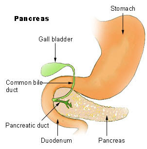
Pancreatitis is the inflammation of the pancreas. The pancreas is an organ anatomically located just underneath our stomach which helps to digest food and also secretes hormones. Among the important hormones secreted are insulin and glucagon which helps to regulate our blood sugar.


Pancreatitis can be divided into acute or chronic pancreatitis depending on the duration and onset of the inflammation.

Patients with Acute Pancreatitis usually present with severe upper abdominal pain which radiates to the back, nausea & vomiting with a history of gallstones or alcohol bingeing. The causes of acute pancreatitis include:
- gallstones
- alcohol ingestion
- trauma (blunt or penetrating) to the abdomen
- post abdominal surgery or ERCP (endoscopic retrograde cholangiopancreaticogram)
- pancreatic tumours or structural abnormalities
- medications such as corticosteroids, diuretics, ACE inhibitors
- autoimmune pancreatitis
- viral infection such as mumps
- high calcium levels
- high fat (triglyceride) levels

Investigations for patients with Acute Pancreatitis include:
- raised serum amylase (usually taken as >4 times normal lab value)
- raised serum lipase
- raised urinary amylase
- raised C-reactive protein
- radiological investigations such as ultrasound or CT scan to look for gallstones or other abnormalities

Patients who are diagnosed with Acute Pancreatitis will need to be admitted into the hospital for close observation. This is because the disease might worsen and lead to severe sepsis and multi-organ failure. The severity of the disease is classified according to the Imrie-Glasgow, Ranson or APACHE II criterias.
Patients are usually kept fasted to reduce the pancreatic function and started on intravenous fluids and also analgesics. They are also given supplemental oxygen.
Patients who have Acute Pancreatitis due to gallstones will need a cholecystectomy (surgical removal of the gallbladder) to avoid further recurrence of the disease.
Patients who are chronic alcoholics should be referred for counselling and also taught coping strategies and encouraged to join Alcoholic Anonymous and other support networks.

The sequelae of Acute Pancreatits include:
- complete recovery (majority of patients)
- destruction and infection of part of the pancreas (necrosis)
- acute fluid collection around the pancreas
- pancreatic pseudocyst (collection of fluid around the pancreas seen >4 weeks after the initial diagnosis)
- pancreatic abscess

Saturday, September 12, 2009

Gallstones

Gallstone disease is common; affecting around 80% of the population. However, of all the patients who have gallstones, only 20% are symptomatic.

There are 3 main types of gallstones:

- Cholesterol stones (80%): commonly occurs in patients who are obese or have high cholesterol and lipids.

- Pigmented stones: Black stones – associated with blood disorders (haemolysis), Brown stones – due to infection of the biliary tract.

- Mixed stones: cholesterol & pigmented types.


Risk factors for gallstones include:

- hereditary

- female sex

- obesity

- high cholesterol levels

- diabetes

- increasing age

- oral contraceptive pills

- post bariartric (obesity) surgery

- inflammatory bowel disease

- haemolytic anaemia


Patients with gallstones can present with:

- abdominal pain usually at the right upper quadrant (biliary colic)

- inflammation of the gallbladder (acute cholecystitis)

- swelling of the gallbladder due to collection of mucous (mucocoele)

- infection of the gallbladder (empyema)

- perforation of the gallbladder (gangrene & perforation)

- jaundice due to obstruction of the bile duct (Mirizzi’s Syndrome)

The gallstones can also migrate into the bile duct & cause obstruction, jaundice & infection.

Common investigations to diagnose gallstones include:

- ultrasound of the abdomen

- CT/MRI scan of the abdomen

- Endoscopic Retrograde Cholangio Pancreaticogram (ERCP): to assess the biliary tract and to remove any stones in the biliary tract.

Treatment:
- Surgery to remove the gallbladder (Cholecystectomy): usually done laparoscopically but can also be done as an open surgery in complicated cases.
- Patients with Acute Cholecystitis will have to be admitted to the hospital for intravenous antibiotics.

- Patients with empyema, gangrenous or perforated gallbladder will need emergency surgery to remove the gallbladder.

Wednesday, September 2, 2009

Thyroglossal Cysts


A Thyroglossal Cyst is a remnant of the thyroglossal tract which connects our tongue to the thyroid gland. During its formation, the thyroid gland bud migrates from the base of our tongue (foramen caecum) to our lower neck. It travels along the thyroglossal tract. The Thyroglossal cyst forms due to the persistent remnant of the thyroglossal tract. Usually the tract disappears completely after the thyroid gland is formed. The tract is closely related to the hyoid bone (a semi-circular bone located just above the thyroid cartilage).
Thyroglossal cysts usually present as a swelling at the middle part of the neck above the thyroid cartilage (adam's apple). However, it can also be located anywhere from the base of the tongue up to just above the sternum (chest bone). It is usually painless but sometimes can get infected. When it gets infected, it might be inflammed & painful. Occasionally the infected thyroglossal cyst might rupture and form a thyroglossal sinus.

It is important to confirm that the swelling is not a lingual thyroid gland prior to surgery. This can be done with an ultrasound scan or a thyroid radioisotope scan.
The treatment for thyroglossal cyst is surgery. This surgery is called Sistrunk's surgery. It involves making a small incision on the cyst and removing it completely together with the remnant tract and also part of the hyoid bone. It is not a major surgery but as always all surgery will carry some risks.

Infected thyroglossal cysts should not be operated on. It should be treated with antibiotics first and planned for a surgery later when the infection subsides.

There is a small risk of malignancy (papillary carcinoma) with the thyroglossal cyst although it is very rare.

Differential diagnosis for a thyroglossal cyst include dermoid cyst, sebaceous cyst, lipoma, lingual thyroid gland and lymph nodes.

Saturday, August 29, 2009

Rhinoplasty (Nose Job Surgery)

Rhinoplasty is one of the commonest cosmetic plastic surgical procedures. It is a nose-reshaping surgery to correct any deformities or to enhance the aesthetics for cosmetic purposes. Rhinoplasty can be done under local or general anaesthesia. The main complaints by patients who are unhappy with their noses are:

- nose bridge hump

- crooked nose bridge

- bulbous nose tip

- long nose tips

- broad & flat nose bridge

- flared nostrils

- post-traumatic injuries to the nose

- birth defects


Our noses can be seen as made up of 3 main components:

1. Nostrils - the two arches of our nose. Some people have bigger & some have narrower arch. Changing the shape of these can change the way the nose looks.

2. Nasal bone & cartilage - these are a collection of cartilages which form the 'nose bridge'. This is where our spectacles/sunglasses rest on. Some people have flatter & wider cartilages and some have higher & narrower ones.

3. Columella - this is the piece of tissue in between our two nostrils. A shorter columella will make the nose look flat while a longer one will make it look higher.


Which part of the nose to be enhanced depends mainly on what the patient wants. Of course, the patients' expectations will have to be reasonable. Rhinoplasty will enhance the patients' looks but will not achieve perfection. The patients should not expect rhinoplasty to transform them into someone else.


It is vital to consult a properly qualified and board certified Plastic Surgeon for rhinoplasty. Other surgeons who do rhinoplasties are ENT & Maxillofacial surgeons.

Wednesday, August 26, 2009

Obesity Surgery

Obesity is defined as a Body Mass Index (BMI) more than 30. Normal BMI is between 18.5 to 25. Patients who have a BMI between 25 to 30 are said to be overweight.

Obesity is associated with many severe medical problems such as hypertension, heart attack, arrhythmias, obstructive sleep apnoea, metabolic syndrome, osteoarthritis, gastroesophageal reflux disease, urinary incontinence, polycystic ovarian syndrome, deep vein thrombosis, depression, low self esteem and increased risk of cancers.
The treatment for obesity includes:

- Lifestyle modifications which includes diet, exercise & behaviour modifications

- Medications. Egs: Sibutramine (appetite suppressant) & Orlistat (reduces fat absorption)

- Surgery:

1. Liposuction - A hollow stainless steel tube (cannula) with the assistance of a powerful vacuum is inserted into the patient’s abdominal skin to suck out the fatty tissue around his/her abdomen. Post liposuction, the patient might get some bruises from the liposuction site which will go away. This is a fast procedure which can be done under general or local anaesthesia. Complications include disfiguring skin irregularities and injury to abdominal organs if not done properly. Liposuction is usually done by qualified Plastic Surgeons only

2. Tummy Tuck (abdominoplasty) - This is a major operation done under general anaesthesia. The Plastic Surgeon will surgically remove a thick layer of fat from the lower abdomen, stretch down the skin from the upper abdomen and then create a new umbilicus for the patient. Post surgery, the patient will need at least 1 week to recover and the abdomen will look flat & trim post-surgery. However, there will be a long surgical scar at the lower waist which can be hidden under the patient’s pants.

3. Bariartric Surgery (Weight Loss Surgery) - a few methods are used but generally divided into "Restrictive" and "Malabsorptive". "Restrictive" surgery reduces the size of your stomach so the end result is you will eat less and then lose weight. The main types are “Gastric Banding”, "Balloon Gastroplasty" or "Sleeve Gastrectomy". "Malabsorptive" surgery is more complicated. Its function is to reduce the amount of intestine available to absorb fat. However, the gold standard is a combination of "restrictive" & "malabsorptive" surgery. Post-surgery, the patient will need at least 5 days to recover depending on the type of surgery. The indications for Bariartric Surgery are:

- Morbid Obesity; which is defined as BMI >40
- Obesity with severe medical conditions
- Failure to lose weight with non-surgical methods
- Medically compliant patient- Patient is medically fit to undergo the surgical procedure

Saturday, August 22, 2009

INFLUENZA A H1N1 - The facts #2

High risk groups:

· Less than 5 years old
· More than 65 years old
· Pregnant
· Obese with chronic illnesses such as asthma, chronic lung disease, heart disease, diabetes or kidney disease
· On long term aspirin therapy
· On drugs that will suppress your immune system eg. steroids, oral chemotherapeutic drugs, azathioprine, cyclophosphamide
· Undergoing cancer treatment


Treatment of H1N1:

Vaccines


Vaccines are usually given to prevent infections. Influenza vaccines are made from either pieces of the killed influenza virus or weakened versions of the live virus that will not lead to disease. When vaccinated, the body’s immune system makes antibodies which will fight off infection if exposure to the virus occurs. However, the influenza virus has the ability to mutate. The latest strain is H1N1. So we will have to wait for the specific vaccine to be available.

Antivirals [Tamiflu (Oseltamivir), Relenza (Zanamivir)]

Antivirals are drugs that can treat people who have already been infected by a virus. Currently, two anti-virals are available which are Tamiflu and Relenza. These drugs are able to stop the virus from replicating in the body and has to be taken within two days after symptoms appear. However, it is not indicated for everybody with flu as the disease is mostly self-limiting. People who are in the high-risk group should be given the anti-virals if they have flu-like illness.
Tamiflu is a neuraminidase inhibitor. It is effective against both type A and B strains of flu.There is no generic version of Tamiflu available. 10 capsules (minimum amount) can cost around RM80 – RM100. Tamiflu helps to reduce your time with flu symptoms by about 1.3 days.

Dosage of Tamiflu:
Oseltamivir is marketed by Roche under the trade name Tamiflu, as capsules (containing oseltamivir phosphate 98.5 mg equivalent to oseltamivir 75 mg). Adults take 75mg twice a day for 5 days.

Side Effects:
The most common side effects of Tamiflu are nausea, vomiting, diarrhea, and conjunctivitis (pinkeye). Sometimes people report getting headaches after taking Tamiflu. There are concerns that oseltamivir may cause dangerous psychological, neuropsychiatric side effects including self harm in some users. These dangerous side effects occur more commonly in children than in adults.


Prevention

The main route of transmission of the new influenza A(H1N1) virus seems to be similar to seasonal influenza, via droplets that are expelled by speaking, sneezing or coughing. You can prevent getting infected by avoiding close contact with people who show influenza-like symptoms (trying to maintain a distance of about 1 metre if possible) and taking the following measures:
- avoid touching your mouth and nose;
- clean hands thoroughly with soap and water, or cleanse them with an alcohol-based hand rub on a regular basis (especially if touching the mouth and nose, or surfaces that are potentially contaminated);
- avoid close contact with people who might be ill;
- reduce the time spent in crowded settings if possible;
- improve airflow in your living space by opening windows;
- practise good health habits including adequate sleep, eating nutritious food, and keeping physically active.


The status of H1N1 now:

The WHO considers the overall severity of the influenza pandemic to be moderate. This assessment is based on scientific evidence available to WHO, as well as input from its Member States on the pandemic's impact on their health systems, and their social and economic functioning.

The moderate assessment reflects that:
- Most people recover from infection without the need for hospitalization or medical care.
- Overall, national levels of severe illness from influenza A(H1N1) appear similar to levels seen during local seasonal influenza periods, although high levels of disease have occurred in some local areas and institutions.
- Overall, hospitals and health care systems in most countries have been able to cope with the numbers of people seeking care, although some facilities and systems have been stressed in some localities.

WHO is concerned about current patterns of serious cases and deaths that are occurring primarily among young persons, including the previously healthy and those with pre-existing medical conditions or pregnancy.
Large outbreaks of disease have not yet been reported in many countries, and the full clinical spectrum of disease is not yet known.

For more info:
Malaysia: http://h1n1.moh.gov.my/
USA: http://flu.gov/
WHO: http://www.who.int/csr/disease/swineflu/en/index.html

Wednesday, August 19, 2009

INFLUENZA A (H1N1) – The Facts

What is it?

H1N1 (previously known as “swine flu”) is a new influenza virus causing illness in people. This new virus was first detected in people in Mexico in April 2009. Now many other countries around the world have reported people sick with this new virus. This virus is spreading from person-to-person, probably in much the same way that regular seasonal influenza viruses spread. This is a new influenza A(H1N1) virus that has never before circulated among humans. This virus is not related to previous or current human seasonal influenza viruses.

History

Over the years, different variations of flu viruses have emerged. At this time, there are four main influenza type A virus subtypes that have been isolated in pigs: H1N1, H1N2, H3N2, and H3N1. However, most of the recently isolated influenza viruses from pigs have been H1N1 viruses.


Signs & symptoms

The symptoms of H1N1 flu in people are similar to the symptoms of regular human flu and include fever, cough, sore throat, body aches, headache, chills and fatigue. Some people have reported diarrhea and vomiting associated with H1N1 infection. Severe illness (pneumonia and respiratory failure) and deaths have been reported with H1N1 (swine) flu infection in people. Like seasonal flu, H1N1 (swine) flu may cause a worsening of underlying chronic medical conditions. You will not be able to tell the difference between seasonal flu and influenza A(H1N1) without medical help. Only your medical practitioner and local health authority can confirm a case of influenza A(H1N1) with a throat swab.


Emergency warning signs in children that need urgent medical attention include:
- Fast breathing or trouble breathing
- Bluish or gray skin color
- Not drinking enough fluids
- Severe or persistent vomiting
- Not waking up or not interacting
- Being so irritable that the child does not want to be held
- Flu-like symptoms improve but then return with fever and worse cough


Emergency warning signs in adults that need urgent medical attention include:
- Difficulty breathing or shortness of breath
- Pain or pressure in the chest or abdomen
- Sudden dizziness
- Confusion
- Severe or persistent vomiting
- Flu-like symptoms improve but then return with fever and worse cough
- Continuous fever for more than three days


Guidelines if you are feeling unwell:
If you feel unwell, have high fever, cough or sore throat:

- stay at home and keep away from work, school or crowds;
- rest and take plenty of fluids;
- cover your nose and mouth when coughing and sneezing and, if using tissues, make sure you dispose of them carefully. Clean your hands immediately after with soap and water or cleanse them with an alcohol-based hand rub;
- if you do not have a tissue close by when you cough or sneeze, cover your mouth as much as possible with the crook of your elbow;
- use a mask to help you contain the spread of droplets when you are around others, but be sure to do so correctly;
- inform family and friends about your illness and try to avoid contact with other people;
- If possible, contact a health professional before traveling to a health facility to discuss whether a medical examination is necessary.
- Do not go to work. You should stay home and away from work through the duration of your symptoms. This is a precaution that can protect your colleagues and others.
- Avoid travelling


For more info:
Malaysia: http://h1n1.moh.gov.my
USA: http://flu.gov/
WHO: http://www.who.int/csr/disease/swineflu/en/index.html

Next: Treatment, prevention & WHO guidance on clinical management of H1N1

Friday, August 14, 2009

Thyroidectomy (Surgery to remove Thyroid Gland)

Thyroidectomy is the medical term for the surgical procedure to remove the thyroid gland. The indications for thyroidectomy are:
- thyroid cancer
- thyrotoxicosis (symptoms of excess of thyroid hormones; refer to earlier topic)
- huge goitre with compressive symptoms (eg. difficulty breathing, difficulty in swallowing)
- cosmesis & patient's request (unsightly neck swelling)


The surgery is performed under general anaesthesia and hospital admission is necessary. Prior to surgery, the patient's thyroid function should be assessed. Patients who are hyperthyroid should be treated with anti-thyroid medications (carbimazole or propylthiouracil) to reduce the levels of thyroid hormones. This is because patients who are hyperthyroid will have an increased risk of a 'thyroid storm'. A thyroid storm is an uncommon but serious complication of thyroid surgery which is precipitated by an acute exacerbation of thyrotoxicosis.


The patient should also undergo an indirect laryngoscopy to assess the patient's vocal cords for any abnormalities. This is because the nerves supplying the vocal cords (recurrent laryngeal nerve) are situated just adjacent to the thyroid gland and any injury will affect the vocal cords. If the goitre is suspicious for cancer, a fine needle aspiration cytology (FNAC) should be carried before surgery to confirm the diagnosis of a cancer. For huge goitres, a chest x-ray or a CT-scan should be done to check for any extension of the thyroid gland inferiorly under the sternum. This would make the surgery more extensive and longer.


After all the pre-operative assesment is done, the patient is admitted for anaesthetic assessment and surgery. During surgery an incision is made just above the collar bones. It is usually around 5-8cm in length depending on the size of the goitre.
There are two options for surgery for thyroid swellings. A hemi-thyroidectomy is performed if the patient has only a single thyroid nodule on one side or small early thyroid cancers (<1cm)>. A total thyroidectomy is performed in patients with a multi-nodular goitre involving both thyroid lobes, thyroid cancers, Graves' disease & thyrotoxicosis. Duration of surgery for a total thyroidectomy ranges from between 1-4 hours depending on the extent of disease and the size of the thyroid gland. Sometimes it might take longer if lymph node dissection is required in cancer patients.

The complications of thyroidectomy can be divided into early and late complications.

Early complications include:
- bleeding
from the surgery wound
- voice change (due to swelling of the nerves)
- temporary low calcium levels (due to reduced function of parathyroid glands)
- wound infection

Late complications include:
- unsightly scar
- permanent low calcium levels (because the parathyroid glands were injured or removed during the surgery)
- permanent hoarseness of voice (injury to recurrent laryngeal nerve)
- vocal cord paralysis with difficulty breathing (injury to both recurrent laryngeal nerves)
- inability to produce a high pitched voice (injury to external branch of superior laryngeal nerve)
- permanent hypothyroidism (in total thyroidectomy, the body cannot produce any more thyroid hormones)

The parathyroid glands are located just behind the thyroid gland in the neck. Usually there are between 2 -6 parathyroid glands in everybody. Its main function is to maintain a normal level of calcium in our body. Due to its location near the thyroid gland, it has a risk of being injured or removed. However, the risk is approximately 1% only. Patients with permanent low calcium levels are treated with calcium supplement tablets.

The recurrent laryngeal nerve supplies our vocal cords. The nerve is located beside the thyroid gland on both sides. The risk of injury is approximately 1%. Injury to the nerve will cause hoarseness of voice (one sided injury) or breathing difficulty (both sided injury). Most patients will have a temporary hoarseness of voice due to swelling of the nerve during manipulation. This resolves spontaneously usually within 1 month.

The external branch of superior laryngeal nerve are located near the upper lobes of the thyroid gland on both sides. Injury to the nerve will lead to an inability to produce high-pitched voice.

Patients who have had a total thyroidectomy will need thyroid hormone supplements for life. This is because there is no more thyroid gland to produce thyroid hormones. This is usually prescribed in the form on levo-thyroxine (L-thyroxine) tablets.

Thursday, August 13, 2009

Health Screening


Medical screening is the process of diagnosing a disease in an otherwise healthy individual before the symptoms occur. The benefits of early diagnosis are that a disease can be treated earlier before it becomes complicated and results in a better outcome. The disadvantages of screening are that there are a small percentage of false positives (diagnosing the disease when in actual fact the patient does not have it), false negatives (missing the diagnosis when the patient actually has the disease) and also the anxiety of being diagnosed with a disease.

There are many different packages for health screening in hospitals and private medical labs. This is what they call "executive health screenings". Some centres also have specially designed packages for women and older people. Usually, a doctor will be on hand for consultation and to get any medical history from you. He/She will then proceed to examine you. Then they will proceed with the investigations such as blood tests, urine tests, chest x-rays, ECGs, Echocardiogram and more.

For routine blood tests, what they check for are:

Full blood count - to check the levels of heamoglobin, platelets and white cells in your body. Basically, this will tell you if you have enough blood in your body (anaemia), any signs of infection and if you have sufficient platelets in your body for clotting.

Renal function test - to check for levels of salt in your body ie. Sodium, Potassium, Chloride. Also to check if your kidneys are functioning well (Serum Creatinine, Urea).

Liver function test - to check if your liver function is normal and if you have enough protein (albumin) in your body. Also can tell if you have jaundice or any liver enzyme/cellular abnormality.

Coagulation profile - to check if your blood clotting mechanism is functioning normally (INR/PT/APTT).

Fasting Blood Glucose – for diagnosis of diabetes mellitus.

Lipid profile – to look at levels of cholesterol (HDL – good /LDL – bad) and triglycerides.

Tumour markers - these are what labs promote the most. Not very sensitive and even if it is raised as it does not mean that the patient has cancer. It is used as a guide in diagnosis of certain cancers. The results should be correlated with clinical findings and with other investigations. Egs: AFP for liver cancer, CEA for colon cancer, CA125 for ovarian cancer, CA19-9 for pancreas cancer, PSA for prostate cancer, Thyroglobulin for thyroid cancer, CA153 for advanced breast and lung cancer.

Blood type and rhesus compatibility - basically telling you your blood type (A/B/O) and whether it is rhesus positive/negative.

ESR/CRP - marker for inflammation. Not very specific.

Uric acid - for diagnosis of gout

Serum Calcium - to check levels of calcium in the body.

Serum T3/T4/TSH - to check levels of thyroid hormones in the body. For diagnosis of hyper or hypothyroidism.

H.Pylori serology – to detect the presence of H.Pylori, a bacteria which causes gastritis and also a risk factor for stomach cancer.

Infectious Disease screen – to check for Hepatitis B, Hepatitis C and HIV.

Urinalysis - to check the urine for protein, bacteria, blood and cells.

ECG (EKG) – to check your heart beat (60 – 100 per minute is normal), heart rhythm (regular or irregular), check for any heart block, check for any angina or ischaemic heart disease.

Echocardiogram – to check if the heart is pumping normally, to look for any problems with the heart muscle, to look at the ventricular function, to look for any heart valvular abnormality.

Mammogram - to detect presence of suspicious breast lumps for diagnosis of breast cancer.